Mediastinal Lymph Node Map
Robin Smithuis
Radiology department of the Alrijne Hospital in Leiderdorp, the Netherlands
Publicationdate
This is an update of the 2007 article, which used the Mountain-Dresler regional lymph node classification for lung cancer staging (MD-ATS maps)(1).
In 2009 a new Lung cancer lymph node map was proposed by the International Association for the Study of Lung Cancer (IASLC) in order to reconcile the differences between the Naruke and the MD-ATS maps and refine the definitions of the anatomic boundaries of each of the lymph node stations (2).
In this article we provide illustrations and CT-images for a better understanding of this IASLC lymph node map.
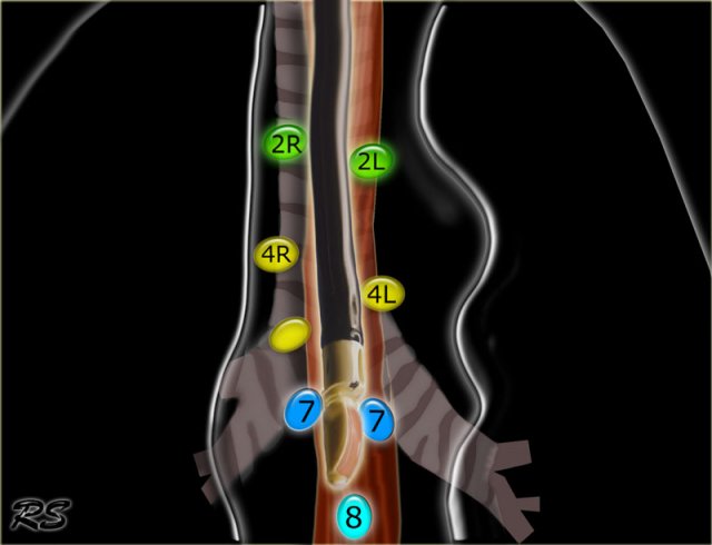
IASLC lymph node map 2009
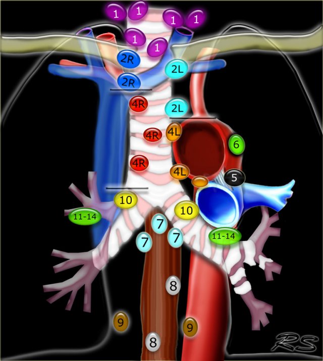 Regional lymph node classification for lung cancer staging adapted from the American Thoracic Society mapping scheme
Supraclavicular nodes
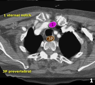
1.Low cervical, supraclavicular and sternal notch nodes
From the lower margin of the cricoid to the clavicles and the upper border of the manubrium.
The midline of the trachea serves as border between 1R and 1L.
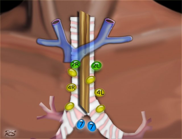
Superior Mediastinal Nodes 2-4
2R.Upper Paratracheal
2R nodes extend to the left lateral border of the trachea.
From upper border of manubrium to the intersection of caudal margin of innominate (left brachiocephalic) vein with the trachea.
2L.Upper Paratracheal
From the upper border of manubrium to the superior border of aortic arch.
2L nodes are located to the left of the left lateral border of the trachea.
3A.Pre-vascular
These nodes are not adjacent to the trachea like the nodes in station 2, but they are anterior to the vessels.
3P.Pre-vertebral
Nodes not adjacent to the trachea like the nodes in station 2, but behind the esophagus, which is prevertebral.
4R.Lower Paratracheal
From the intersection of the caudal margin of innominate (left brachiocephalic) vein with the trachea to the lower border of the azygos vein.
4R nodes extend from the right to the left lateral border of the trachea.
4L.Lower Paratracheal
From the upper margin of the aortic arch to the upper rim of the left main pulmonary artery.
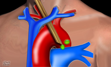
Aortic Nodes 5-6
5. Subaortic
These nodes are located in the AP window lateral to the ligamentum arteriosum.
These nodes are not located between the aorta and the pulmonary trunk but lateral to these vessels.
6. Para-aortic
These are ascending aorta or phrenic nodes lying anterior and lateral to the ascending aorta and the aortic arch.
Inferior Mediastinal Nodes 7-9
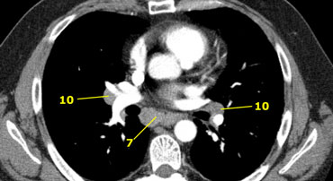
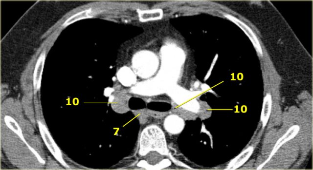
7.Subcarinal
8. Paraesophageal
Nodes below carina.
9. Pulmonary Ligament
Nodes lying within the pulmonary ligaments.
Hilar, Lobar and (sub)segmental Nodes 10-14
These are all N1-nodes.
10. Hilar nodes
These include nodes adjacent to the main stem bronchus and hilar vessels.
On the right they extend from the lower rim of the azygos vein to the interlobar region.
On the left from the upper rim of the pulmonary artery to the interlobar region.
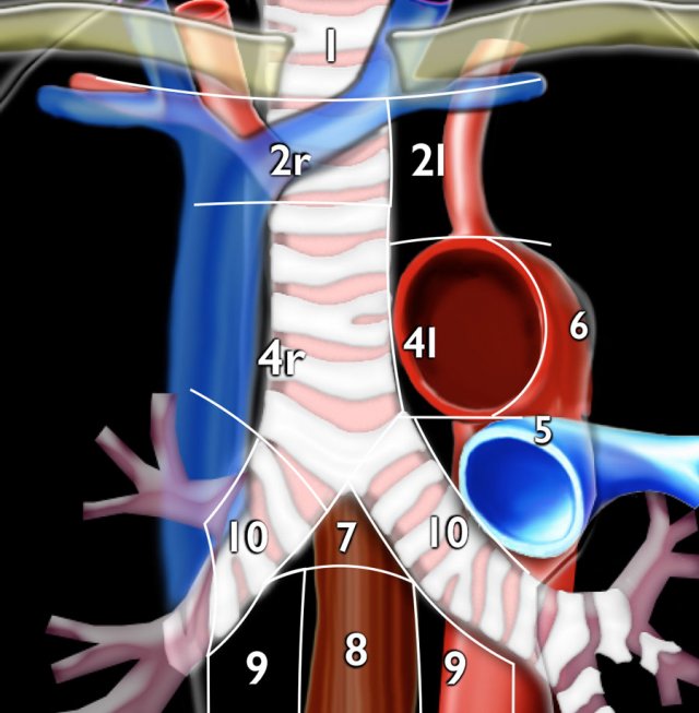
Axial CT anatomy
Click on image to enlarge.
Then scroll through the axial CT-images.
Images by Dr. Aurelia Fairise of the Institut de Cancérologie de Lorraine in Nancy.
Specific Lymph Node Stations
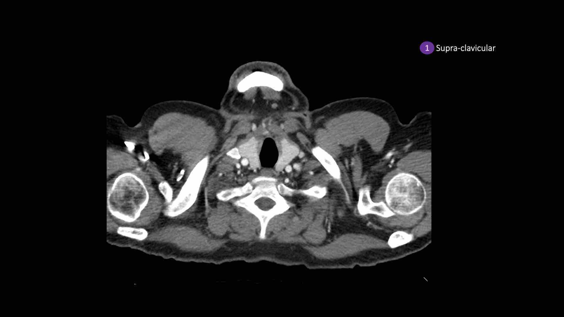
 1. Supraclavicular zone nodes
1. Supraclavicular zone nodes
These include low cervical, supraclavicular and sternal notch nodes.
Upper border: lower margin of cricoid.
Lower border: clavicles and upper border of manubrium.
The midline of the trachea serves as border between 1R and 1L.
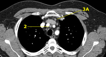
2R. Right Upper Paratracheal
2R nodes extend to the left lateral border of the trachea.
Upper border: upper border of manubrium.
Lower border: intersection of caudal margin of innominate (left brachiocephalic) vein with the trachea.
2L. Left Upper Paratracheal
Upper border: upper border of manubrium.
Lower border: superior border of aortic arch.
On the left a station 2 node in front of the trachea, i.e. a 2R-node.
There is also a small prevascular node, i.e. a station 3A node.
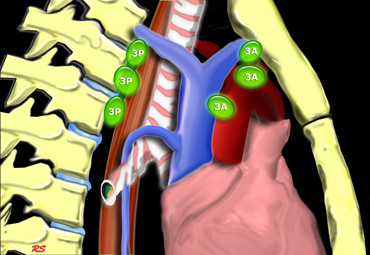 3A and 3P nodes
3. Prevascular and Prevertabral nodes
Station 3 nodes are not adjacent to the trachea like station 2 nodes.
They are either:
3A anterior to the vessels or
3B behind the esophagus, which lies prevertebrally.
Station 3 nodes are not accessible with mediastinoscopy.
3P nodes can be accessible with endoscopic ultrasound (EUS).
On the left a 3A node in the prevascular space.
Notice also lower paratracheal nodes on the right, i.e. 4R nodes.
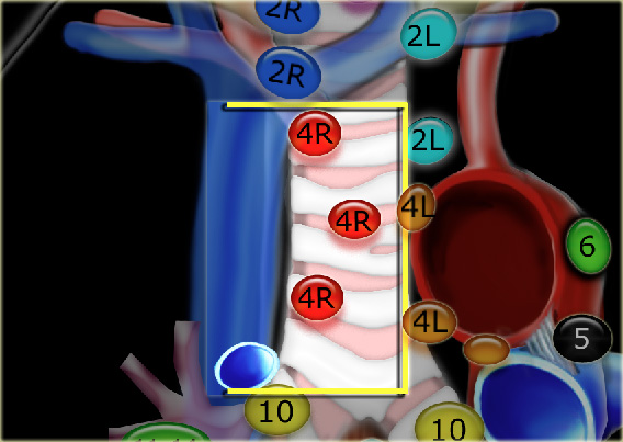 4R. Lower Paratracheal nodes
4R. Right Lower Paratracheal
Upper border: intersection of caudal margin of innominate (left brachiocephalic) vein with the trachea.
Lower border:lower border of azygos vein.
4R nodes extend to the left lateral border of the trachea.
On the left we see 4R paratracheal nodes.
In addition there is an aortic node lateral to the aortic arch, i.e. station 6 node.
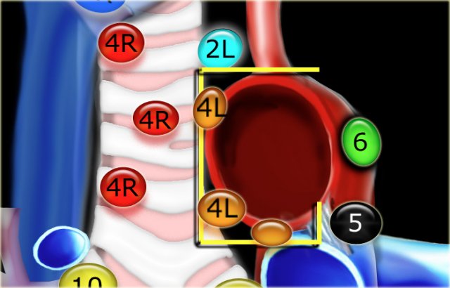 4L. Lower paratracheal nodes
4L. Left Lower Paratracheal
4L nodes are lower paratracheal nodes that are located to the left of the left tracheal border, between a horizontal line drawn tangentially to the upper margin of the aortic arch and a line drawn tangentially to the upper margin of the left pulmonary artery.
These include paratracheal nodes that are located medially to the ligamentum arteriosum.
Station 5 (AP-window) nodes are located laterally to the ligamentum arteriosum.
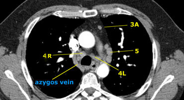
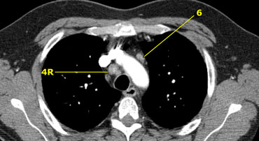
On the left an image just above the level of the pulmonary trunk demonstrating lower paratracheal nodes on the left and on the right.
In addition there are also station 3 and 5 nodes.
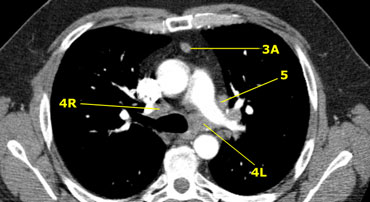
On the left an image at the level of the lower trachea just above the carina.
To the left of the trachea 4L nodes.
Notice that these 4L nodes are between the pulmonary trunk and the aorta, but are not located in the AP-window, because they lie medially to the ligamentum arteriosum.
The node lateral to the pulmonary trunk is a station 5 node.
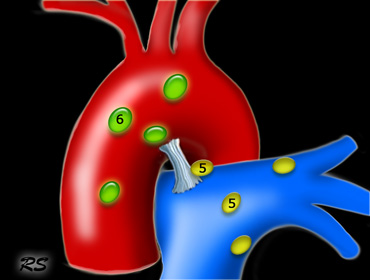
5. Subaortic nodes
Subaortic or aorto-pulmonary window nodes are lateral to the ligamentum arteriosum or the aorta or left pulmonary artery and proximal to the first branch of the left pulmonary artery and
lie within the mediastinal pleural envelope.
6. Para-aortic nodes
Para-aortic (ascending aorta or phrenic) nodes are located anteriorly and laterally to the ascending aorta and the aortic arch from the upper margin to the lower margin of the aortic arch.
7. Subcarinal nodes
These nodes are located caudally to the carina of the trachea, but are not associated with the lower lobe bronchi or arteries within the lung.
On the right they extend caudally to the lower border of the bronchus intermedius.
On the left they extend caudally to the upper border of the lower lobe bronchus.
On the left a station 7 subcarinal node to the right of the esophagus.
 .
.
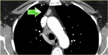
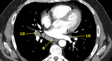
8 Paraesophageal nodes
These nodes are below the carinal nodes and extend caudally to the diafragm.
On the left an image below the carina.
To the right of the esophagus a station 8 node.
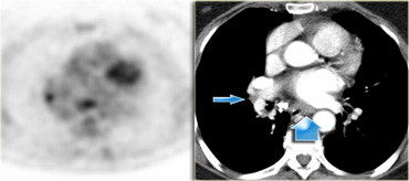
On the left a PET image demonstrating FDG uptake in a station 8 node.
On the corresponding CT image the node is not enlarged (blue arrow).
The probability that this is a lymph node metastasis is extremely high since the specificity of PET in unenlarged nodes is higher than in enlarged nodes.
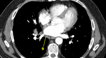
9. Pulmonary ligament nodes
Pulmonary ligament nodes are lying within the pulmonary ligament, including those in the posterior wall and lower part of the inferior pulmonary vein.
The pulmonary ligament is the inferior extension of the mediastinal pleural reflections that surround the hila.
10 Hilar nodes
Hilar nodes are proximal lobar nodes, distal to the mediastinal pleural reflection and nodes adjacent to the intermediate bronchus on the right.
Nodes in station 10 - 14 are all N1-nodes, since they are not located in the mediastinum.
Axial CT of Lymph Nodes
Scroll through the images on the left.
- Sternal notch nodes are just seen at this level and above this level
- Upper Paratracheal: below clavicles and on the right above the intersection of caudal margin of innominate (left brachiocephalic) vein with the trachea and on the left above the aortic arch.
- Pre-vascular and Retrotracheal : anterior to the vessels (3A) or prevertebral (3P)
- Lower Paratracheal : below upper margin of aortic arch down to level of main bronchus
- Subaortic (A-P window): nodes lateral to ligamentum arteriosum or lateral to aorta or left pulmonary artery
- Para-aortic: nodes lying anterior and lateral to the ascending aorta and the aortic arch beneath the upper margin of the aortic arch
- Subcarinal
- Paraesophageal (below carina)
- Pulmonary Ligament: nodes lying within the pulmonary ligament.
- -14: nodes are all N1 nodes
Mediastinoscopy and EUS
Conventional mediastinoscopy
The following nodal stations can be biopsied by cervical mediastinoscopy: the left and right upper paratracheal nodes (station 2L and 2R), left and right lower paratracheal nodes (station 4L and 4R) and the subcarinal nodes (station 7).
Station 1 nodes are located above the suprasternal notch and are not routinely accessed by cervical mediastinoscopy.
Extended mediastinoscopy
Left upper lobe tumors may metastasize to the subaortic lymph nodes (station 5) and paraaortic nodes (station 6).
These nodes can not be biopsied through routine cervical mediastinoscopy.
Extended mediastinoscopy is an alternative for the anterior-second interspace mediastinotomy which is more commonly used for exploration of mediastinal nodal stations.
This procedure is far less easy and therefore less routinely performed than conventional mediastinoscopy.
EUS-FNA
Endoscopic Ultrasound with Fine Needle Aspiration can be performed of all the mediastinal nodes that that can be assessed from the oesophagus.
In addition the left adrenal gland and the left liver lobe can be visualized.
EUS particularly provides access to nodes in the lower mediastinum (station 7,8 and 9)